
CellChek CC20
Gold Standard Specular Microscopy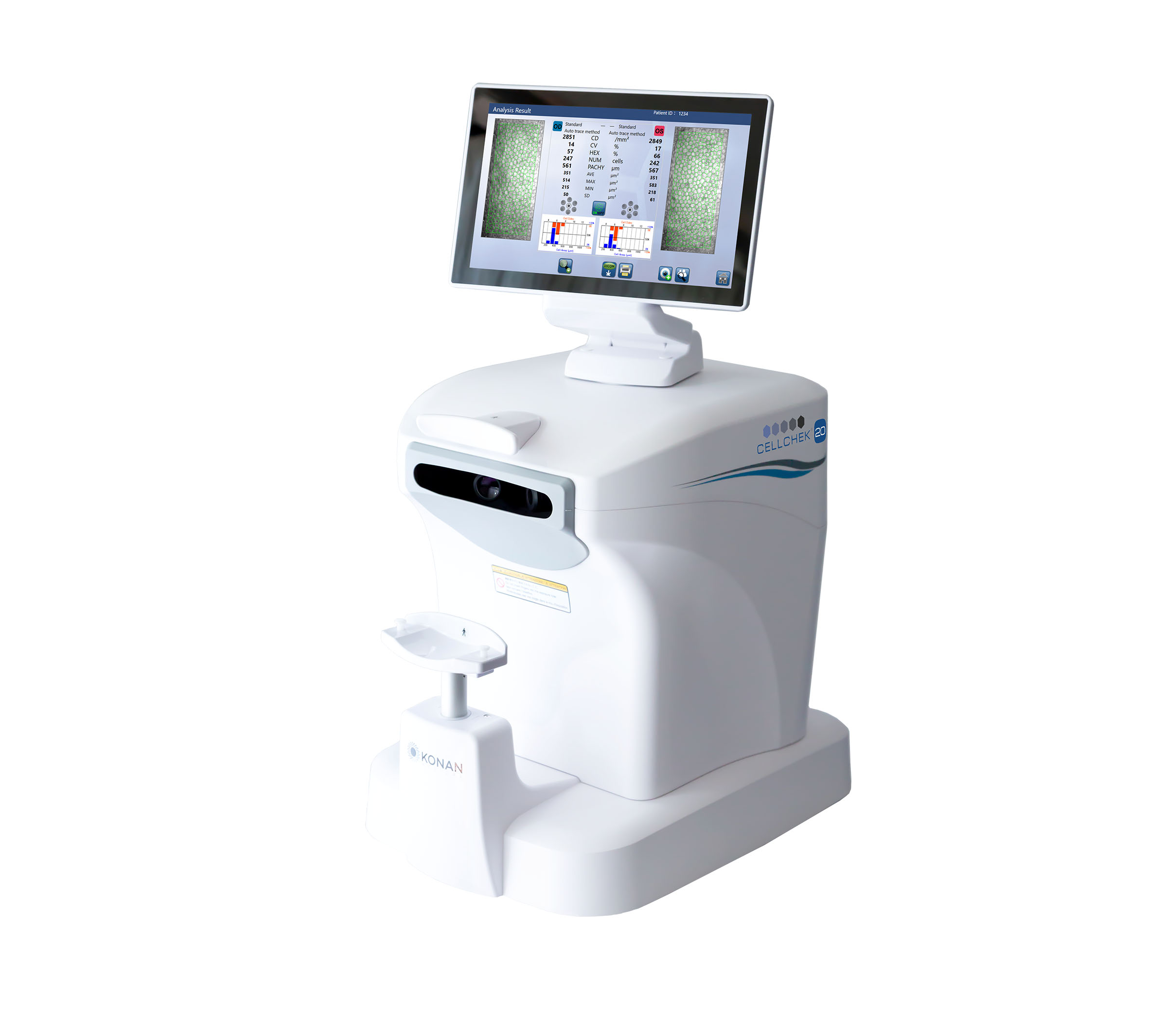
Low endothelial cell counts and pre-existing dystrophies can markedly reduce the potential for positive surgical outcomes from an otherwise uneventful cataract surgery.
In assessing the success of corneal graft surgery, knowing the pre- and post-op cell counts and being able to view any changes graphically over time, gives the surgeon rapid and valuable data to help manage patients post-surgery.
It is not a coincidence … that virtually every manufacturer of pharmaceutical agents and implantable devices that are required by the FDA to collect endothelial cell density as a primary safety endpoint, have had their clinical research teams, completely independently, assess the specular microscopes available on the market and have chosen Konan. Repeatedly.
CC-20 Fully Automated Specular Microscope
The Gold Standard, redefined, for faster, easier endothelial Imaging
CellChek 20 is Konan Medical’s new, non-contact specular microscope that can capture and analyse bilateral exams with one touch, in under 40 seconds. The new easy-to-use “Simple Mode” enables one-touch, fully-automated endothelial image capture with analysis, reporting, and exporting of data.
Flexible Touch-Screen
The flexible 10.6” wide touch screen can be turned and tilted 180°, providing better ergonomics, space savings, and flexibility with technician use from any of the four sides: front, back, left, and right.

Fully-Automated Analysis
CellChek 20 offers fully-automated: Centre Method, Flex-Centre Method and Auto-Trace analysis. These fully-automated analysis methods are offered alongside the traditional manual capability, providing a total of 6 analysis options. Konan’s Centre Method is mentioned in FDA panel minutes as being the “gold standard” and is used by virtually every professional reading centre.

New Auto Centre Method
With the new Auto Centre Method, the centre of each individual visible cell, is automatically marked. Guttae and other dark regions are automatically excluded.
New Auto Flex-Centre Method
The new Auto Flex-Centre Method is an additional automated tool for analysing images of advanced stage diseased corneas in which only a very few cells are visible.
